- Who is a Candidate for Nuclear Medicine Breast Imaging?
- How is Nuclear Medicine Breast Imaging Performed?
- Recent Research on Nuclear Medicine Breast Imaging
In a recent study conducted by Italian researchers, mammography and nuclear medicine breast imaging were compared in 134 women aged 32 to 78. While the overall accuracy of the two tests were similar, mammography was less likely to identify breast cancer in the younger women than the nuclear medicine test. This suggests that nuclear medicine may be effective in women with dense breast tissue. The researchers concluded that nuclear medicine may help in surgical planning because of its high specificity and could be considered complimentary to mammography, especially in younger women. A Turkish study also found that nuclear medicine breast imaging may be helpful in detecting breast cancer that had spread to the axillary (armpit) lymph nodes. In fact, nuclear medicine imaging is sometimes used with sentinel lymph node biopsy to help determine if the lymph nodes contain cancer cells.
In another study by researchers from the Los Robles Regional Medical Center in California, nuclear medicine breast imaging was evaluated in 75 patients with signs on either mammography or physical exam that might or might not have indicated breast cancer. Of the 30 diagnosed cancers, 27 were positively identified with nuclear medicine. Eight of those 27 cancers were not identified with mammography or physical exam, and 11 of the cancers were smaller than one centimeter. The researchers concluded that nuclear medicine is a useful method of evaluating patients with indeterminate (difficult to read) mammograms or physical exams and may help detect additional small breast tumors. However, further research is needed to confirm the results of this study, especially since previous studies have shown that nuclear medicine may not be helpful in detecting small breast abnormalities.
Click here to learn more about recent research on nuclear medicine breast imaging.
Case #1:
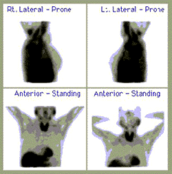
This nuclear medicine study shows normal breast anatomy and no nodules in this patient:
 |
Case #2:
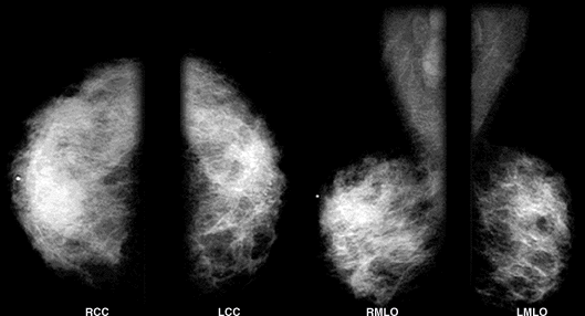
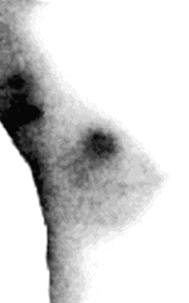
In the following case of a 40-year old woman, a physician-performed clinical breast exam revealed a mass in the right breast. The mammogram images showed extremely dense breasts and no suspicious masses or calcifications. However, the technetium sestamibi image showed focal uptake in the right breast in the area of palpable mass. A breast biopsy was then performed and the pathology results showed infiltrating ductal carcinoma, a type of breast cancer.
The patient’s mammogram shows extremely dense breasts and no suspicious masses or calcifications:
 |
This right lateral technetium sestamibi image shows focal uptake in the right breast in the area of the palpable (able to be felt) mass. Image courtesy of Miraluma:
 |
- The September 19, 2001 Imaginis Report, "Nuclear Medicine Breast Cancer Test May Be Helpful for Some Women, Particularly in Those with Dense Breasts" is available at http://www.imaginis.com/breasthealth/news/news9.19.01.asp
- General information about nuclear medicine imaging is available at http://www.imaginis.com/nuclear-medicine/
- To learn more about how breast cancer is diagnosed, please visit http://www.imaginis.com/breasthealth/menu-diagnosis.asp
Updated: May 4, 2008


