- What is a Sentinel Lymph Node?
- What is Sentinel Lymph Node Biopsy?
- How is Sentinel Lymph Node Biopsy Performed?
- Why is Sentinel Lymph Node Biopsy Performed?
- What Happens When the Sentinel Node Is Found To Be Cancerous?
- What Are the Side Effects of a Sentinel Lymph Node Biopsy?
- Is Sentinel Node Biopsy Accurate?
- Are All Breast Cancer Patients Candidates for Sentinel Lymph Node Biopsy?
- Additional Resources and References
 |
|
Courtesy
of Tyco |
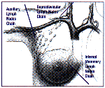
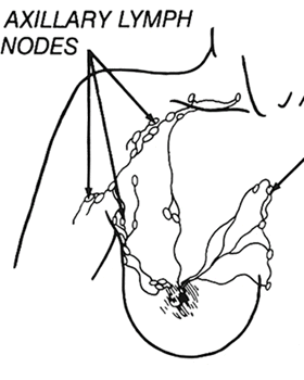
In the breast, a network of
lymphatic vessels drain fluid and cells to the bean-shaped lymph nodes in the axilla
(armpit). The sentinel node is the very first lymph node(s) to receive
drainage from a cancer-containing area of the breast.
Put another way, when breast cancer cells begin to escape from the primary tumor site in
the breast they travel to the lymph nodes under the arm; the first lymph node they reach
is the 'sentinel' lymph node.
When breast cancer is diagnosed,
women (and men) must often undergo axillary lymph node
dissection (i.e., removal of underarm nodes) to check for the spread of cancer. This
process is part of staging the cancer.
Unfortunately, the removal of these lymph nodes can lead to lymphedema (chronic
swelling) of the arm in a certain percentage-- about 10-20%--of cases.
Sentinel lymph node biopsy is a new diagnostic procedure used to determine whether breast
cancer has spread (metastasized) to axillary lymph
nodes (i.e., lymph glands under the arm). A sentinel lymph node biopsy requires the
removal of only one to three lymph nodes for close review by a pathologist. If the
sentinel nodes do not contain tumor (cancer) cells, this may eliminate the need to remove
additional lymph nodes in the axillary area.
Early research on this technique indicates that sentinel lymph node biopsy may be
associated with less pain and fewer complications than standard axillary dissection. However, because the
procedure is so new, long term data are not yet available.
Before going to the operating room, the surgeon injects a small dose of a low-level radioactive tracer called technetium-99 into the breast in the region of the patient's tumor. Technetium-99 contains less radiation than a standard x-ray, CT scan or bone scan and is a relatively safe substance. A blue dye is also injected to help visually track the location of the sentinel node during surgery. The surgeon then uses a hand held counter to detect the radioactive tracer and locate the sentinel node.
Sometimes, nuclear medicine images (also known as lymphoscintigraphy) of the lymphatic system will be obtained after injecting the technetium-99 before surgery. Since the uptake of the technetium-99 by cancerous lymph nodes is sometimes different than the uptake by normal lymph nodes, these nuclear medicine images may also help show which lymph nodes are cancerous.
Next, the surgeon will wait for the technetium-99 and dye to travel from the tumor region to the sentinel lymph node(s), just as cancer cells might spread. Depending on the protocol followed, the surgeon usually waits between 45 minutes to 8 hours after injection before bringing a patient to the operating room for the biopsy. At some point during the procedure, a small amount of blue dye will also be injected into the breast tissue near the area of the tumor. Once the technetium-99 tracer and dye have reached the nodes, the surgeon will scan the area with an electric, hand-held gamma ray counter (called a Geiger counter) to detect the radioactive technetium-99.
The gamma ray counter is attached to a small probe which the surgeon traces over the axilla to locate the sentinel node(s). When the radioactive agent is found, the gamma ray counter will emit an audible tone, revealing the exact location of the sentinel node(s). Once the area has been pinpointed, the surgeon will make a small incision (usually one-half inch) and remove the sentinel node(s) for a pathologist to examine under a microscope. The blue dye provides additional visual confirmation of the sentinel node’s location during surgical removal. Several clinical trials have revealed that in the vast majority of cases, if the sentinel node does not contain cancer, then the cancer has not spread past the breast. Sentinel node biopsy does not usually require the placement of a fluid drainage tube (common in axillary node dissection).
 |
|
Courtesy NIH/NCI |
Sentinel lymph node biopsy may help in
determining which patients can avoid axillary node dissection and the removal of 10 to 30
lymph nodes. Most patients have only one to three sentinel lymph nodes under the
arm. Thus, an average of only two lymph nodes are removed in each patient with a
sentinel node biopsy. This, in turn, may reduce post-operative complications.
A standard axillary node dissection, removal of the
underarm lymph nodes, usually requires a larger four to six inch incision and a longer
recovery period than a sentinel node biopsy.
Researchers are currently investigating whether sentinel node biopsy should routinely be
performed in place of axillary node dissection. However, surgeons caution that more
studies on the benefits and risks of sentinel node biopsy should be conducted before the
procedure becomes widespread. Typically, patients who undergo a modified radical mastectomy or a lumpectomy may
require lymph node removal. Sentinel node biopsy or axillary node dissection helps
surgeons determine if breast cancer has spread to the lymphatics and the extent of the
spread.
In a recent study published in The New England Journal of Medicine, 443 patients at 11 medical centers across the United States underwent sentinel node biopsies. Researchers discovered that if the gamma ray counter detected the radioactive agent (technetium-99) in the patients, then the sentinel node biopsy was 97% accurate in pinpointing all cancerous lymph nodes. However, the study was not completely successful. The gamma ray counter missed cancerous nodes in 13 of the 114 women whose cancer had spread past the breast. In an upcoming study, researchers at the National Cancer Institute will compare sentinel node biopsy to the standard method of lymph node removal (axillary node dissection) on 4000 women to determine which procedure is superior.


