- Who Is/Is Not a Candidate for Lumpectomy?
- How is Lumpectomy Performed?
- Radiation Therapy After Lumpectomy
 |
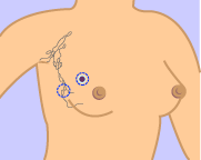
This illustration shows how lumpectomy is performed by removing the tumor and margin of surrounding normal breast tissue. Some of the axillary (underarm) lymph nodes may also be removed in patients who undergo lumpectomy. Illustration courtesy of the NCI/NIH. |
Lumpectomy may be performed using a local anesthetic, sedation, or general anesthesia, depending on the extent of the surgery needed. The surgeon makes a small incision over or near the breast tumor and excises (cuts free) the lump or abnormality along with a margin of at least one centimeter (approximately one half inch) of normal surrounding breast tissue (see the section above for information on margins). Unlike after mastectomy, a drainage tube is usually not necessary after lumpectomy.
A seroma (clear fluid trapped in the wound) usually fills the surgical cavity after the operation and helps to naturally remold the breast’s shape. Gradually, the seroma is absorbed and the body replaces it with scar tissue. This natural healing process and formation of scar tissue occurs over a period of months, so that the final results of the surgery may not be apparent for some time. Depending on such factors as the location of the mass, its initial size, the type of incision used, etc., the final result will be different for each person.
| Possible Side Effects of Lumpectomy Include: |
|
Patients are usually able to go home the same day or one to two days following lumpectomy. Most women are able to resume normal activities within two weeks. Wound infection or bleeding is not common with lumpectomy. The extent of breast soreness correlates with the amount and location of tissue removed during surgery, whether axillary (underarm) lymph node surgery was performed, and an individual’s tolerance to pain. Major soreness usually ceases after two to three days and should be checked by a physician if there is any increase in pain over time. Because lumpectomy is usually intended to preserve the cosmetic appearance of the breast, surgeons generally do not recommend lumpectomy when over one fourth of the breast must be removed. In these cases, mastectomy, along with the option of reconstruction, may be preferable.
In rare instances, women may experience recurring seromas after lumpectomy. Seromas are collections of fluid in the cavity (empty space) left behind by the surgery. These collections are easily drained (aspirated) in the surgeon’s office. If a seroma recurs, surgeons may use several methods including compression or sclerosis (the injection of ethanol, autologus fibrin clot, or fibrin sealant) to fill and harden the space in the breast. At times, these treatments can be uncomfortable, but they are rarely needed.
Lumpectomy (and sometimes mastectomy) is typically followed by six to seven weeks of radiation therapy immediately following surgery to help ensure that any remaining cancer cells are destroyed and to help prevent the chance of a cancer recurrence. Treatment with radiation usually begins one month after surgery, allowing the breast tissue adequate time to heal. Treatments are given daily and each treatment generally lasts a few minutes; the entire radiation session after machine set-up typically lasts 15 to 30 minutes. The procedure itself is pain-free. While the radiation is being administered, the technologist will leave the room to monitor the patient on a closed-circuit television. However, patients should be able to communicate with the technologist at any time over an intercom system.
| Common Side Effects of Radiation Therapy |
|
Most of the side effects associated with radiation therapy are temporary, and many patients do not experience significant discomfort after radiation sessions.
Researchers have been investigating whether a shorter duration, higher dose of radiation may be as effective as the conventional six to seven week regimen. Recent research suggests that limiting radiation therapy to four weeks at a higher dose may be as effective as the traditional regimen and could reduce side effects. IMRT (intensity-modulated radiation therapy) uses a highly sophisticated system of delivering external-beam radiation. According to recent research, this system uses advanced computer optimized planning and radiation delivery techniques that create more optimal dose distributions, greater sparing of the skin and lower doses to organs such as lung and heart--thus reducing potential side effects. However, there may be patients who are uncomfortable with the idea of an accelerated treatment and want to be treated with a more conventional six to seven week course of treatment. "In addition, we need more research to determine which women are ideal candidates for this treatment because of differences in anatomy or other treatments for their breast cancer." Women are encouraged to talk to their cancer treatment team about their radiation therapy options.
Click here for more information on radiation therapy.
 |
|
|
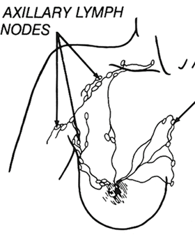
When breast cancer cells begin to escape from the primary tumor site in the breast, they first travel to the lymph nodes under the upper arm. Therefore, it is often necessary to remove some or all of the axillary (underarm) lymph nodes during lumpectomy or mastectomy to determine if or to what extent the cancer has spread.
Lymph node removal usually requires a separate incision when it is performed during the same procedure as lumpectomy. There are two procedures for removing lymph nodes in breast cancer patients: axillary node dissection and sentinel node biopsy.
- Axillary node dissection: This is the standard way to remove axillary lymph nodes. Typically, between 10 to 30 lymph nodes are removed and examined in a pathology laboratory to determine whether they contain cancer cells.
- Sentinel lymph node biopsy: This is a technique that involves the injection of a blue dye, radioactive tracer, or both, to identify the "sentinel" lymph nodes (first nodes) draining the breast. Using this method, only the first one to three lymph nodes in the lymphatic chain are removed. Research has shown that checking the sentinel lymph nodes allows physicians to accurately determine whether the axillary (armpit) lymph nodes contains cancer while causing fewer side effects such as lymphedema (chronic swelling) of the arm. If the sentinel nodes are positive (contain cancer cells), then additional surgery is performed to remove (dissect) the remaining axillary lymph nodes. If the removed axillary lymph nodes are negative (do not contain cancer cells), then no additional lymph nodes are removed, reducing the side effects of axillary dissection. Sentinel lymph node biopsy has become more common in recent years. However, it is not always appropriate. Click here for more information about this procedure.
The most common side effect of lymph node removal is lymphedema (chronic swelling) of the arm. Between 10% and 20% of patients who have lymph nodes remove develop lymphedema, including some patients who only have a sentinel lymph node biopsy. The risk of lymphedema is greater if the patient also undergoes radiation therapy and/or the lymph nodes contained cancer cells upon final examination. To help manage lymphedema and prevent long-term suffering, patients should report symptoms as soon as they occur. In addition, special exercises should be performed shortly after recovering from surgery to help encourage and maintain lymphatic flow of the affected side of surgery.
|
Early Signs of Lymphedema |
|
In addition to lymphedema, other common side effects of lymph node removal include limitations of arm/shoulder movement, and numbness of the upper arm skin. Click here to learn more about lymphedema.
- The American Cancer Society, http://www.cancer.org, provides detailed information on lumpectomy and other breast cancer treatment options.
- To learn more about lymphedema, please visit http://www.imaginis.com/breasthealth/lymphedema.asp
Updated: April 2012


