 |
The technologist uses the hand-held T-scan probe to image the breast. Image courtesy of Siemens Medical Solutions. |
The T-scan imaging system is similar in size and appearance to an ultrasound system. There is a small cart-mounted unit with a monitor and keyboard. The patient holds a metallic wand that is similar in size and appearance to a "joy-stick" on many computer games. One to two and a half volts (1.0V to 2.5V) of compatible alternating (AC) electrical current are generated by the system and conducted through the body from the wand held by the patient. When the scanning probe is placed against the skin of the breast the electrical circuit is completed. Gel is used as an agent to improve conductivity between the skin of the breast and the scanning probe (similar to ultrasound). A minuscule amount of electric current is used, approximately the same amount produced by a small penlight battery. The procedure creates no discomfort, and in most patients, no sensation.
The scanning probe is moved over the breast and its many sensors measures the current signal at the skin level. The computer then reconstructs this information and shows images immediately on the monitor. The scanning probe uses 256 sensors in high-resolution mode and 64 sensors in normal-resolution mode. Images of the electrical impedance profile of the breast are produced in real-time and are displayed as a 256-level gray scale map of capacitance and conductivity properties of the breast. The image is recorded into 9 sectors on a 3 x 3-sector matrix electrical impedance map of the breast. Targeted electrical impedance views may be recorded using the anatomic screen.
Impedance objects are defined as spots or regions that are brighter (or occasionally darker) than their surrounding sector image. The only white object that should occur in T-scan examination of a normal breast is the nipple. Skin marks such as moles, insect bites, or recent biopsy locations can also show as bright spots and must be annotated by the operator so they are not confused with cancerous abnormalities. Under normal conditions, the nipple image should be the same size and symmetrical for both breasts.
|
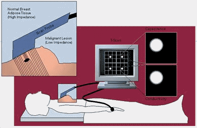
| This figure shows how the scanning
probe is held against the breast and measures the low level current that is conducted
through the breast tissue in the region of interest. Normal tissue has a high impedance
(low conductivity), malignant (cancerous) tissue has a lower impedance (higher
conductivity). The measured signals are then digitally processed by the system's computer
and displayed immediately as an image on the monitor. Image courtesy of Siemens Medical Solutions, www.medical.siemens.com |
Cancer cells have lower electrical impedance than healthy cells. When the T-scan signal is displayed on the computer screen, possible tumors show up as bright white spots. Cytological (cellular) and histological (tissue) changes in cancerous tissue, including changes in cellular water and electrolyte content and cell membrane properties, cause a significant change in tissue electrical impedance (20-40 times lower than normal tissue), enabling cancerous and pre-cancerous lesions to be visualized.
Updated: January 27, 2000


