Before radiation treatment is started, x-ray films are taken and calculations are made to determine the angles from which the x-rays should be directed. These measurements are taken with a simulation machine, either an x-ray simulator or a CT simulator. Sometimes both simulators are used.
During simulation, the area of the patient's body to be treated is marked directly on the patient's skin with markers. Very tiny permanent marks, or tattoos are sometimes made to ensure that daily treatments are delivered accurately. These are also useful for future treatment planning sessions and treatment updates.

A radiation oncology physicist plans a treatment session using CT data
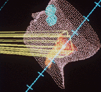
In a growing number of radiation therapy centers, radiation physicists and dosimetrists use computer models to plan the treatment. This computerized treatment planning allows the treatment to be delivered more accurately, both in terms of the "beam shape" that is achieved with precise collimator settings and in terms of the angles and directions used to deliver the radiation to the cancer. By using the computer to help "focus the x-ray beam," the radiation oncologist can deliver the radiation precisely to cancer, delivering a minimal amount of radiation to healthy tissues.
The radiation treatment planning models usually use a three-dimensional (3D) re-creation of the patient's anatomy. Images of the patient are acquired from the CT or MR scanner and then sent by network or computer disk to the radiation treatment planning computer.

Computer model of the path energy rays will take during radiotherapy of a brain tumor
Updated: June 10, 2008


