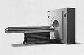
Positron Emission Tomography (PET) scanning is a type of nuclear medicine scanning that involves cross sectional data acquisition and reconstruction much like Computed Tomography (CT) scanning. PET scanning has specific potential in imaging certain diseases and disorders of the brain (for example brain tumors) and the heart. Lung cancer imaging is a new, emerging application of PET and involves inhalation of the radionuclide, rather than oral or intravenous administration.

This PET image shows a significant number of "hot spots" throughout the patient's body
- Study of epilepsy (nervous system disorders that cause convulsive seizures)
- Evaluation of stroke (blood clot or bleeding in the brain)
- Study of dementia (for example in patients with Alzheimer's or Parkinson's disease)
- Imaging and evaluation of brain tumors
- Evaluation of coronary artery disease and detection of transient ischemia (poor blood flow)
- Differentiation between recurrent, active tumor growth and necrotic (dead) soft tissue masses in cancer treatment patients

SPECT (single positron emission computed tomography) is another type of nuclear medicine examination. SPECT uses a gamma camera which can rotate, and computer reconstruction similar to PET. In some cases, PET may be more sensitive than SPECT, but PET scanners are much more costly than SPECT scanners and are often only available in the largest medical centers.
Updated: June 10, 2008


