Magnetic Resonance (MR or MRI) is the preferred method of imaging the brain to detect the presence of plaques or scarring caused by multiple sclerosis (MS). Due to the excellent tissue contrast of MR, it is better at detecting plaques than computed tomography (CT or CAT scanning). Often, brains that appear to be normal on CT images will be shown to have MS plaques on MR images.

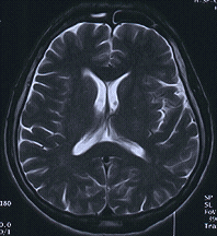
Axial T2 weighted MR image, bright CSF obscures the MS plaque in this brain, even using high resolution matrix

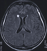
Fluid-suppressed "Turbo FLAIR" MR image, the bright MS plaque is now apparent. Images courtesy of Siemens Medical Solutions
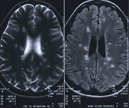
MR became clinically available in the mid-1980s. Traditionally, a "T2" MR image of the brain was used to show MS lesions (areas of damage or disease) and to diagnose multiple sclerosis. T2 MR images show brain tissue as dark and cerebrospinal fluid (CSF) as bright. MS lesions are also show as bright areas or spots on T2 images. In some cases, the bright CSF fluid made the identification of MS plaques difficult on standard T2 images. However, newer MR techniques (sequences) called "dark fluid" or "Turbo FLAIR" have been developed to suppress the bright CSF on T2 images so that the MS plaques are more evident. These "fluid suppressed T2" sequences are available on advanced MR systems. These sequences can be performed at various magnetic field strengths including lower field strength magnets used with "Open MR" systems.

MS diagnosis with advanced Open MR system. T2 image on the left and Turbo-FLAIR image on the right. Image courtesy of Siemens Medical Solutions.
The diagnosis of MS cannot be made solely on the basis of MR imaging. There are other neurologic diseases such as Glioma that cause lesions in the brain that appear similar to MS plaques on MR images. There are also abnormalities found in healthy individuals, particularly in older persons, that are not related to any ongoing disease process.
On the other hand, a normal MR does not absolutely rule out a diagnosis of MS. Approximately five percent of patients who are confirmed to have multiple sclerosis on the basis of other criteria, do not show any plaques or lesions in the brain on MR images. These people may have lesions in the spinal cord or may have plaques that cannot be detected by MR imaging.


