A breast abscess is a closed pocket of tissue containing pus (a creamy, thick, pale yellow or yellow-green fluid). Abscesses are most commonly caused by a bacterial infection. Abscesses may or may not show up well on ultrasound. Breast abscesses may be accompanied by fever, pain, breast tenderness, or increased white blood cell count.
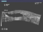
This panoramic ultrasound image shows full visualization of a breast abscess.
Fibroadenomas are common benign (non-cancerous) breast tumors often too small to feel by hand, though occasionally, they may grow to be several inches in diameter. Fibroadenomas are made up of both glandular and stromal (connective) breast tissue and usually occur in women between 20-30 years of age. Fibroadenomas often stop growing or even shrink on their own without any treatment. In these cases, doctors do not recommend having the tumors removed. Fibroadenoma surgery may involve removing a margin of breast tissue surrounding the fibroadenoma.
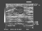
Image shows circumscribed, slightly lobulated fibroadenoma.
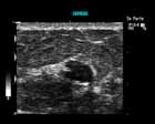
Image of fibroadenoma.
A breast mass is any group of breast cells that are clustered together more densely than the surrounding breast tissue. Masses can be palpable (able to be felt) or nonpalpable (unable to be felt). Masses can be benign or cancerous.
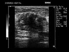
This image shows a solid irregular breast mass with calcification (calcium deposits). Ultrasound does not reliably image calcifications, although they can be seen in some cases.
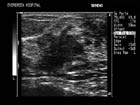
This image shows ductal invasion associated with this malignant breast mass.
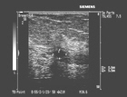
This image shows a nonpalpable (unable to be felt) mass within the glandular breast tissue.


