


- How Does the CAD Technology Work?
- Studies with CAD Technology
- Benefits and Limitations of CAD Technology
Computer-aided detection (CAD) technology is a recent advance in the field of breast imaging. The CAD technology basically works like a second pair of eyes, reviewing a patient's mammogram film after the radiologist has already made an initial interpretation. If the computer software detects any breast abnormalities or "regions of interest" on the mammogram film, it marks them. The radiologist can then go back and review the mammogram film again to determine whether the marked areas are suspicious and require further examination (with additional imaging tests or biopsy). With the CAD technology, the radiologist still makes the final interpretation of the mammogram.
Based on clinical studies of the CAD technology, researchers estimate that for every 100,000 breast cancers currently detected with screening mammograms, the CAD technology could result in the detection of an additional 20,500 breast cancers.
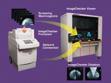
Photo courtesy of R2 Technology, Inc.
Mammography can help detect breast cancer at an early stage, when the chances for successful treatment and survival are the greatest. While mammography detects approximately 85% to 90% of breast cancers, mammogram films can be difficult for radiologists to read. Thus, radiologists can occasionally overlook breast cancers that are present on mammogram films. The CAD technology works as a "second reading" for radiologists, alerting them to areas on films that may require more attention.
Recently, Congress passed the "Medicare, Medicaid, and SCHIP Benefits Improvement and Protection Act of 2000" that will provide additional reimbursement for screening mammograms using the CAD technology. According to California Representative Anna Eschoo, this legislation will give women greater access to more accurate mammograms.
In essence, the CAD technology works like a "spell-checker." The computer marks abnormalities on the digitized films similar to the way a computer program might alert a writer to a misspelled word. After reviewing the computer’s marking, the radiologist can decide whether the marked area is indeed an abnormality that needs follow-up or if the computer has alerted him or her to a normal area, such as a blood vessel, that is no cause for concern. The final interpretation is still made by the radiologist.
To use the CAD technology, mammogram films are first loaded into a special processing unit that digitizes the mammogram images. The CAD unit then highlights any detected breast abnormalities on the digitized mammograms using special pattern recognition computer software. The digitized mammogram files are then transmitted to monitors on a motorized film viewer so the radiologist can compare the original film to the digitized mammogram image on the small monitor.
In the meantime, the radiologist reviews the patient’s original mammogram films and makes his or her interpretation as to whether any breast abnormalities are present (and whether they are cause for concern). After the radiologist finishes analyzing the mammogram films, he or she can view the digitized mammograms on the small monitor to determine whether the computer marked any abnormalities on the films. Based on the results of the CAD marker information, the radiologist may choose to re-examine the original mammogram films and modify his or her interpretation when appropriate.
Using sophisticated pattern recognition computer software, the CAD technology is designed to detect the following abnormalities on mammogram films:
- Patterns of bright spots that suggest microcalcifications (tiny calcium deposits that may indicate cancer)
- Dense regions with or without radiating lines that suggest breast masses or distortions
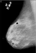 |
The CAD system marks suspicious areas on the digitized mammogram film. Photos courtesy of R2 Technology, Inc. |
The CAD technology marks breast abnormalities on digitized mammography films using a special coding system. For example, the R2 Imagechecker marks clusters of calcifications with a small triangle and breast masses with an asterisk. (See the mammogram image below). CAD marks are only made on the digitized mammograms; the original films are not altered.
