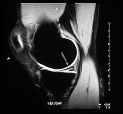
Injury or disease is physically manifested by actual change in a person's anatomy. For example, if a person has a cartilage tear of his or her knee from a sports injury, MR (MRI) imaging can clearly show this damage. An MR image would depict a tear of the knee's meniscus (knee joint's surface) as a white mark on the meniscus. A healthy meniscus normally appears as a small, totally black triangle on specific types of MR images. The white mark shown is typically fluid that has collected in the tiny tear.

MR image of the knee, side view (sagittal)
showing damaged meniscus
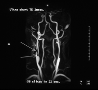
Or, if a person has a small plaque (blockage caused by cholesterol build up in the interior vessel wall), an angiogram will depict this disease as a small restriction or lump inside the vessel.

MR angiogram of the carotid (neck)
arteries showing restricted vessel
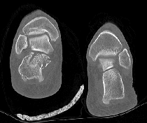
A broken bone can also be seen on an conventional x-ray or CT scan, the break often appears just as one would imagine it would look like. However, subtle fractures can be very painful and often required a well trained radiologist to diagnose.

CT image of the ankles showing fracture; note the
small pieces of fractured bone on the left
Each of the five main diagnostic imaging modalities
has certain strengths and weaknesses in depicting various diseases or injuries. In many cases, two or more of the five main modalities can be used in combination to provide the physician with the most complete information.
Interpreting diagnostic images, however, is not as simple as it sounds. It is a skilled science and an art. Well trained and experienced radiologists can often immediately see pathology that would be altogether missed by a lay person and would take a less skilled physician a long while to visualize. For more information, see the section on "Who are the professionals that perform diagnostic imaging?"
Updated: August 2010


