


Computed Tomography (CT) imaging, also known as "CAT scanning" (Computed Axial Tomography), combines the use of a digital computer together with a rotating x-ray device to create detailed cross sectional images or "slices" of the different organs and body parts such as the lungs, liver, kidneys, pancreas, pelvis, extremities, brain, spine, and blood vessels. For many patients, CT can be performed on an outpatient basis without requiring admittance to the hospital.
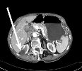
Axial CT image of the liver and kidneys shows a benign (non-cancerous) cyst in the right kidney (arrow)
Among the various imaging techniques such as MR and x-ray, CT has the unique ability to image a combination of soft tissue, bone, and blood vessels. For example, conventional x-ray imaging of the head can only show the dense bone structures of the skull. X-ray angiography of the head only depicts the blood vessels of the head and neck and not the soft brain-tissue. Magnetic resonance (MR) imaging does an excellent job of showing soft tissue and blood vessels, but MR does not give as much detail of bony structures such as the skull. CT images of the head allow physicians to see soft-tissue anatomic structures like the brain's ventricles or gray and white matter. Physician then can selectively "window" the digital CT images on the computer monitor to look at the soft tissue, then the bone and then the blood vessels, as needed.
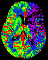
Colored CT image of the brain, purple area on right of image indicates acute stroke
