


- Main Menu:
- Breast Biopsy Overview
- Digital Spot-View Mammography Improves Breast Biopsy
- Breast Biopsy with Ultrasound Image Guidance
- Breast Biopsy with Prone (Face Down) Stereotactic Image Guidance
- Breast Biopsy with Upright Stereotactic Image Guidance
- Relative Cost of Different Breast Biopsy Methods
- Breast Biopsy Overview
- Digital Spot-View Mammography Improves Breast Biopsy
- Breast Biopsy With Ultrasound Image Guidance
- Breast Biopsy With Prone (Face Down) Stereotactic Image Guidance
- Breast Biopsy With Upright Stereotactic Image Guidance
- Relative Costs of Different Breast Biopsy Methods
- Methods of Breast Biopsy
- Benefits and Risks to Breast Biopsy
A breast biopsy involves removing a sample of breast tissue to determine whether it is cancerous or benign (non-cancerous). There are several different types of breast biopsy. The biopsy method most suitable for a particular patient depends on a number of factors, including:
- whether or not an abnormality can be felt or only seen with imaging
- how suspicious the abnormality appears on x-ray (or feels on palpation)
- the size, shape, and other distinct characteristics of the abnormality
- the location of the abnormality in the breast and in relation to other anatomic structures
- the number of abnormalities detected during physical examination or with x-ray imaging
- the patient’s medical history and current medications
- the preference of the patient, as long as an option is medically safe and appropriate
In addition, the systems available at a given breast imaging center or surgical center may influence which method of biopsy a patient receives. For example, vacuum-assisted biopsy (brand name, Mammotome or MIBB) is currently not available at all facilities.
Many biopsy methods rely on image guidance to help the radiologist or breast surgeon precisely locate the lesion (abnormality) within the breast. Imaging may be necessary when a lesion (breast abnormality) cannot be felt during examination and is only detected on imaging studies such as mammography or ultrasound. It may also be necessary to use imaging to assure that a mass felt during examination is, indeed, the same abnormality noted on a mammogram or ultrasound.
If image guidance is required, each of the factors listed above will also be considered in terms of what type of image guidance is most appropriate for the biopsy. Biopsy methods that may require image guidance include:
- fine needle aspiration biopsy (FNA)
- core needle biopsy
- vacuum-assisted biopsy (Mammotome or MIBB)
- large core biopsy (ABBI)
- open surgical biopsy (excisional or incisional)
Of these methods, FNA, core needle, and vacuum-assisted biopsies are performed as percutaneous ("through the skin") procedures rather than surgical biopsies. Click on one of the biopsy methods listed above for a detailed explanation of the procedure.
Though percutaneous ("through the skin") biopsy methods only provide partial information for pathologists, they are usually very accurate when performed by a skilled radiologist or surgeon who has significant experience with image guidance.
However, because percutaneous biospies only provide samples of tissue (and not the entire lesion as a surgical biopsy does), occasionally, additional procedures may be needed for final diagnosis and treatment of a breast abnormality. This is especially true if there are faint calcifications (calcium deposits), small or low density masses, or diffuse (spread out) abnormalities. Diffuse lesions can be difficult to locate accurately in two stereotactic x-ray views.
Therefore, women should always discuss the risks and benefits of any recommended procedure with their physicians before deciding on the most suitable diagnostic option. Click here to learn more about the different methods of breast biopsy.
A recent advance in the field of mammography is digital mammography. Digital (computerized) mammography is similar to standard mammography except that breast images are obtained using a system that is equipped with a digital receptor and a computer instead of a film cassette. Digital spot view mammography is often used during breast biopsy and typically allows a faster and more accurate stereotactic biopsy. This usually results in a shorter procedure, increasing patient comfort.
With digital spot-view mammography, images are acquired digitally and displayed immediately on the system monitor. Traditional stereotactic biopsy requires a mammogram film be exposed, developed and then reviewed, significantly increasing the time before the breast biopsy can be completed. Today, the majority of facilities use digital mammography when image guidance is needed for stereotactic biopsy.
Fine needle aspiration (FNA), core needle biopsy, and vacuum-assisted biopsy may be performed using ultrasound (or stereotactic image guidance) if a breast lesion is easily visualized with this method.
Ultrasound is an excellent method of imaging the breasts; ultrasound guidance allows biopsy of the breast from almost any orientation.
During an ultrasound-guided biopsy, small samples of tissue are removed from the breast using a hollow core needle or vacuum-assisted biopsy device that is precisely guided to the correct location using continuous ultrasound imaging. During the procedure, the patient will lie face up on the standard ultrasound table while being modestly draped, exposing only the area of the breast undergoing biopsy. The area is identified by a preliminary ultrasound.
During a prone stereotactic biopsy, the patient lies face down (prone) on a specially designed table with the breast placed through an opening in the tabletop. "Stereotactic" means that the breast biopsy path is imaged from two slightly angled directions to help guide the needle. The tabletop is raised and the radiologist and technologist perform the procedure from beneath. The patient's breast is slightly compressed and held in position throughout the procedure. Several stereotactic pairs of x-ray images are made. Small samples of tissue are then removed from the breast using a hollow core needle or vacuum-assisted biopsy device that is precisely guided to the correct location using x-ray imaging and computer coordinates.
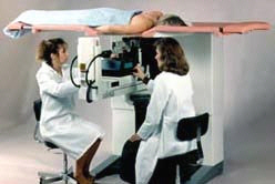 |
The radiologist and technologist
perform a prone stereotactic biopsy. |
Patients who weigh over 300 pounds are usually not appropriate candidates for biopsies that require the use of the prone table. These patients may be better candidates for upright stereotactic image guidance biopsies.
Breast biopsy using upright stereotactic mammography requires the patient to sit upright in a chair or to lie on her side (lateral recumbent position). The breast biopsy path is imaged from two slightly angled directions to help guide the needle using an upright stereotactic mammography system. The patient’s breast is slightly compressed and held in position throughout the procedure. Several stereotactic pairs of x-ray images are made. Small samples of tissue are then removed from the breast using a hollow core needle or vacuum-assisted biopsy device that is precisely guided to the correct location using x-ray imaging and computer coordinates. Upright biopsy is the best method for guiding a biopsy of the axilla or "tail" of the breast; 41% of breast cancers occur in this region.
Perhaps the most difficult part of a biopsy with upright stereotactic image guidance for many patients is the need to sit upright without moving during the biopsy. Therefore, upright stereotactic mammography may not be a viable option for some elderly, anxious, or physically impaired persons. However, upright stereotactic mammography may be an appropriate method for patients who would find it difficult to climb up onto the prone table.
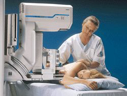 |
The patient is in the lateral recumbent position for biopsy using an upright stereotactic mammography system. Image courtesy of Siemens Medical. |
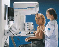 |
The patient is in the seated position during biopsy using an upright stereotactic mammography system. Image courtesy of Siemens Medical. |
Many health insurance providers cover the cost of a breast biopsy. However, women should always check with their insurance companies before undergoing any medical procedure to determine whether the cost of the biopsy and related costs will be covered (and at what rate). Women should also check to see if special referrals are necessary, as many health maintenance plans require these prior to any specialist consultations or procedures.
The cost of a breast biopsy can range from approximately $1000 to approximately $5000 depending several factors, including the type of biopsy performed, the equipment used during the biopsy, whether image guidance or other additional equipment is necessary to perform the biopsy, etc. The cost of a breast biopsy typically increases as the procedure becomes more invasive. This is due to increases in associated costs of using technologically advanced equipment and forms of anesthesia. The type of facility where the biopsy is performed may also influence the cost. For example, the cost may be higher if the biopsy is performed in an outpatient suite at a hospital versus an outpatient surgical center. Fine needle aspiration (FNA) is usually the least expensive biopsy method while open surgical biopsy tends to be the most expensive.
|
Average Expense for Breast Biopsy |
| 1. Fine
Needle Aspiration (FNA) - least expensive 2. Core Needle 3. Vacuum-Assisted (Mammotome or MIBB) 4. Large Core (ABBI) 5. Open Surgical - most expensive |
Updated: May 4, 2008
